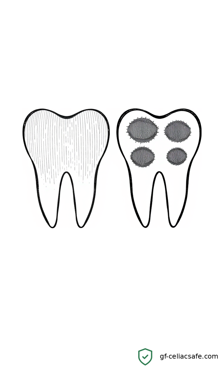
Children with celiac disease are at higher risk for a specific kind of dental defect — soft, chalky enamel on permanent molars and front teeth that decays fast and causes chronic pain. New research published in Frontiers in Nutrition suggests this defect is not random chance. It connects directly to how much gut damage a child has sustained and how depleted their fat-soluble vitamins are.
The study, by Cristun, Vasilescu, and Mang, examined children with celiac disease for a condition called molar incisor hypomineralisation (MIH) — patches of poorly formed enamel on the first permanent molars and incisors. Children with MIH had lower levels of fat-soluble vitamins (A, D, E, and K) and higher markers of intestinal damage — how “leaky” the gut lining had become — compared to celiac children without the defect.
For families managing pediatric celiac disease, this is significant. Dental enamel does not grow back. The window for forming healthy permanent teeth closes by roughly age eight. If celiac disease goes undiagnosed or is poorly managed during those early years, the nutritional gaps during that window can leave permanent marks on a child’s teeth.
As a parent of a child with celiac disease, I read this study and immediately thought about the families I hear from who struggle with overlapping health issues — dental problems, poor growth, fatigue — that seem unrelated until you look at the underlying nutritional picture. This research draws that connection more clearly than most studies I have come across.
What This Means for You
The most important takeaway here is that dental enamel defects in a celiac child are a clinical signal, not a coincidence.
If your celiac child has MIH — chalky white or brown spots on permanent molars or incisors, teeth that break or decay unusually fast — that pattern may reflect vitamin deficiency during early tooth development. That is worth discussing with your child’s gastroenterologist and dentist together. The dental finding is a window into nutritional history.
Fat-soluble vitamins are absorbed in the small intestine, the same part of the gut that celiac disease damages. When the intestinal lining is inflamed and permeable, absorption of vitamins A, D, E, and K drops. Vitamin D is especially critical for tooth mineralization — it regulates how calcium and phosphorus get incorporated into developing enamel. A deficiency during the years when permanent teeth are forming can produce exactly the kind of soft, defective enamel this study describes.
We have covered the broader nutritional picture in pediatric celiac disease before — in Nutritional status in pediatric celiac disease: evaluation at diagnosis and during follow-up, we discussed how deficiencies often persist even after diagnosis and the transition to a gluten-free diet. This new study adds a concrete, visible consequence of those deficiencies: teeth that bear lasting damage from a gap in care that may have closed long before the diagnosis was made.
The gut permeability angle is equally important. A leakier gut means poorer nutrient absorption overall — not just lower vitamin levels on a lab report, but functionally less of those vitamins reaching the developing tissues that need them. Managing celiac disease aggressively — strict gluten-free diet, regular monitoring, addressing nutritional gaps — matters not just for gut health but for the whole body, including teeth.
Key Takeaways
- Children with celiac disease have higher rates of MIH, a developmental defect leaving permanent molars and front teeth with soft, poorly formed enamel.
- This study found that celiac children with MIH had lower fat-soluble vitamin levels (A, D, E, K) and a leakier gut lining than celiac children without it.
- Vitamin D deficiency during early childhood — a common consequence of undiagnosed or undertreated celiac disease — impairs enamel formation.
- Dental enamel defects in a celiac child are worth investigating as a nutritional signal, not dismissing as a separate problem.
- Strict adherence to the gluten-free diet and proactive vitamin monitoring may help protect teeth in children diagnosed early.
The Science
Want to understand how this actually works? We’ll walk you through the technical details below and define every term. No medical degree required.
What Is Molar Incisor Hypomineralisation?
Molar incisor hypomineralisation (MIH) is a developmental defect in which the enamel of the first permanent molars — and often the upper front teeth — fails to mineralize properly during formation. The result is enamel that is structurally weak. It may appear chalky white, yellow, or brown, and it breaks down rapidly under normal chewing forces.
MIH is not cavities in the traditional sense. It is a formation defect that happens before the teeth ever erupt into the mouth. Once it exists, it cannot be reversed. Management options are limited to protective sealants, restorations, and in severe cases, extraction.
Celiac disease has long been recognized as a cause of dental enamel defects — one of its most common extra-intestinal (outside-the-gut) manifestations. What this study adds is a mechanistic explanation: the link runs through fat-soluble vitamins and gut barrier integrity, and the severity of the dental defect tracks with how severe those underlying problems are.
The Role of Fat-Soluble Vitamins in Tooth Development
Fat-soluble vitamins — A, D, E, and K — differ from water-soluble vitamins like C and B12 in how they are absorbed. They require dietary fat and a functioning small intestine. In celiac disease, the villi (tiny finger-like projections lining the small intestine that absorb nutrients) are flattened and damaged by the immune response to gluten. This dramatically reduces fat-soluble vitamin absorption.
Of these vitamins, vitamin D is most directly tied to tooth mineralization. It regulates how the body uses calcium and phosphorus — the minerals that give enamel its hardness. During enamel formation, specialized cells called ameloblasts deposit these minerals in a precise crystal structure. Without adequate vitamin D, this process fails. The enamel that forms is softer, less organized, and far more vulnerable.
Vitamin A plays a supporting role. It governs the differentiation and function of ameloblasts. A deficiency disrupts the orderly formation of the enamel matrix — the protein scaffold that mineralization depends on. Together, deficiencies in these vitamins during the first several years of life can set a child up for irreversible dental problems before anyone suspects celiac disease is involved.
Intestinal Permeability as a Marker
Intestinal permeability refers to how readily substances pass through the gut wall. A healthy gut maintains tight junctions between cells — essentially sealed seams that prevent large molecules, toxins, and antigens from passing into the bloodstream. In active celiac disease, these junctions loosen, and the gut becomes more permeable — often called “leaky gut” in lay terms.
Researchers frequently measure this using markers like zonulin, a protein that regulates the tight junctions between intestinal cells. Higher zonulin levels indicate a more permeable gut. This study found that higher permeability correlated with worse dental outcomes in celiac children — suggesting that the degree of gut barrier compromise tracks with the severity of downstream nutritional consequences, including those that affect developing teeth.
This builds on a growing body of research examining how body composition and nutritional deficits reflect the extent of celiac-related gut damage — including earlier work on Bioelectrical Impedance Analysis to Identify the Affected Body Component in Girls With Celiac Disease, which identified measurable body-composition differences in girls with the condition.
What Still Needs to Be Studied
This research establishes association, not definitive causation. We know MIH and vitamin deficiency co-occur in celiac children, but the precise timing and dose-response relationship between nutritional status and enamel damage still needs prospective study. It is also not yet clear whether aggressive early supplementation of fat-soluble vitamins in newly diagnosed celiac children can meaningfully reduce MIH rates going forward.
These are not trivial questions. Enamel defects are irreversible. If early nutritional intervention can protect teeth, that is a compelling reason to act before symptoms appear — and well before the window for enamel formation closes.
Consult your child’s gastroenterologist and pediatric dentist about monitoring fat-soluble vitamin levels and dental development if your child has been diagnosed with celiac disease. This article is informational and does not constitute medical advice.
Related Coverage
- Nutritional status in pediatric celiac disease: evaluation at diagnosis and during follow-up
- Bioelectrical Impedance Analysis to Identify the Affected Body Component in Girls With Celiac Disease
References
- Cristun LI, Vasilescu RN, Mang N. Molar incisor hypomineralisation in relation to fat-soluble vitamin status and intestinal permeability in children with celiac disease. Frontiers in Nutrition. 2026. PubMed