A new case report raises an important diagnostic warning. A rare variant of the blistering skin rash classically linked to celiac disease can look nearly identical to the standard form—but often has no connection to celiac at all. Published in the Journal of Cutaneous Pathology, the paper describes two patients with this atypical presentation and warns that failing to recognize it can put patients on gluten-free diets they don’t need.
The condition is dermatitis herpetiformis (DH), sometimes called “celiac of the skin.” In the vast majority of cases, DH is a celiac disease manifestation and responds to a strict gluten-free diet. But researchers Damman, Mendels, and van der Woude identified two patients whose DH looked different at the microscopic level—different enough that neither patient had any evidence of celiac disease at all.
What This Means for You
For celiac families, DH is a familiar part of the landscape. We’ve covered how common DH is among celiac patients and the neurological complications it can carry. This new report is about something different: a DH lookalike that can be confused with the classic form and mistakenly attributed to celiac disease.
The practical risk cuts in two directions. Classic DH is treated primarily with a gluten-free diet. If a patient actually has this rare fibrillar variant—which the authors say is often unrelated to celiac—they could be told to go gluten-free without any genuine need. A lifelong gluten-free diet is not a minor inconvenience. It’s expensive, socially limiting, and demands constant vigilance. Prescribing it without a confirmed celiac connection is a real harm to a patient’s quality of life.
The other risk runs the opposite way. A clinician unfamiliar with fibrillar DH might dismiss an atypical presentation entirely when it doesn’t match the textbook mold, leaving a patient without a correct diagnosis or appropriate treatment for whatever is actually causing their rash.
This is a case report involving two patients, not a large clinical trial. It won’t rewrite diagnostic guidelines overnight. But it adds to a growing body of evidence that DH is more varied than the standard description suggests—and it reinforces a principle that celiac families know well: thorough diagnostic workups matter before any major intervention is prescribed.
If someone in your family is being evaluated for DH or celiac disease based on a skin rash, this research is worth raising with their care team. It’s another reason to ask whether the diagnostic picture is complete.
Key Takeaways
- Dermatitis herpetiformis (DH) is the intensely itchy blistering rash classically linked to celiac disease—but a rare variant called fibrillar DH often has no celiac connection.
- Two patients in this case report had fibrillar DH with no evidence of celiac disease.
- Classic DH shows granular immune deposits in the skin; fibrillar DH shows thread-like deposits and lacks the celiac-linked skin enzyme entirely.
- Misidentifying fibrillar DH as classic DH can result in patients being placed on gluten-free diets they don’t need.
- Clinicians need to assess the shape and structure of immune deposits—not just their presence—before concluding a patient has celiac-related DH.
The Science
Want to understand how this actually works? We’ll walk you through the technical details below and define every term. No medical degree required.
How Classic Dermatitis Herpetiformis Works
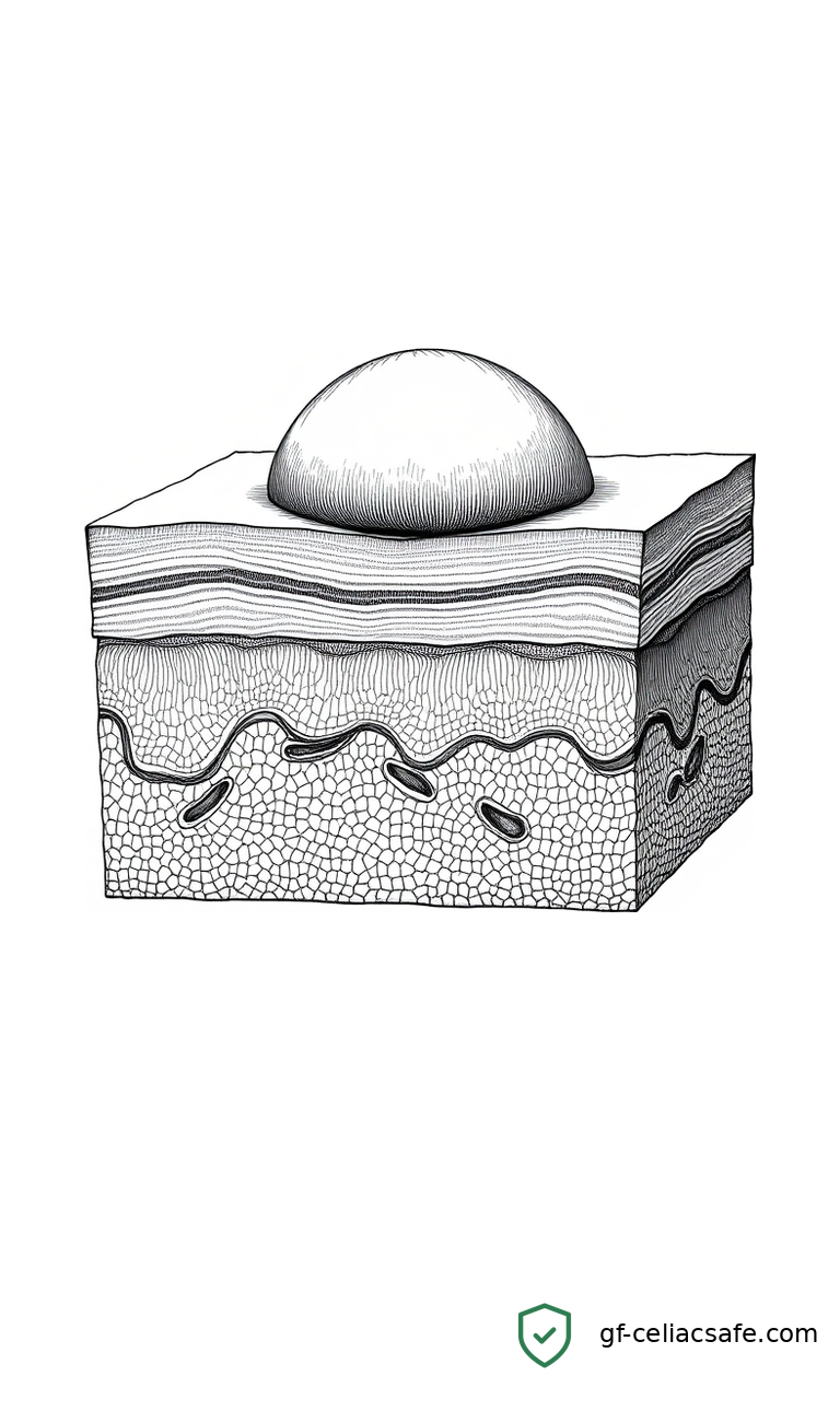
In classic DH, the immune system produces IgA autoantibodies—proteins that mistakenly attack the body’s own tissue. These autoantibodies specifically target an enzyme in the skin called epidermal transglutaminase (eTG). The antibodies accumulate in the uppermost layer of skin tissue, called the papillary dermis, forming immune deposits that trigger intense itching and subepidermal blistering.
Under a microscope using a technique called direct immunofluorescence (DIF)—which uses fluorescent tags to reveal immune deposits—those deposits appear granular: small, distinct clumps of immune material clustered near the skin surface. This granular IgA pattern is the diagnostic standard for DH. When a dermatologist or pathologist sees it, celiac disease is almost always the underlying cause.
What Sets the Fibrillar Variant Apart
In both patients described in this report, the IgA deposits looked completely different. Rather than granular clumps, the deposits appeared fibrillar—arranged in thin, thread-like strands. This fibrillar pattern has appeared in prior case literature, but it remains rare and poorly characterized.
The more significant finding is what the researchers confirmed for the first time: eTG did not colocalize with the fibrillar IgA deposits. Colocalization means two substances appear in the same location in tissue—in this case, the researchers used specialized imaging to determine whether eTG and IgA overlapped in the skin samples. In classic DH, they overlap, which explains why the immune response targets that specific enzyme. In these two patients, they didn’t overlap at all.
That absence is diagnostically meaningful. If eTG isn’t part of the immune complex driving the skin condition, the underlying mechanism differs from classic DH. The fibrillar IgA deposits may be binding to a different target, or they may represent a distinct disease process that simply resembles DH when tissue samples are examined.
Why This Creates a Diagnostic Risk
Standard skin biopsies detect the presence of IgA deposits, but routine lab analysis may not distinguish fibrillar from granular patterns without careful attention to morphology—the shape and structure of the deposits. A clinician who sees IgA in the papillary dermis might reasonably assume classic DH, and from there, assume celiac disease. Neither assumption holds when the pattern is fibrillar.
The authors argue that DIF results need to be interpreted carefully, and that the specific morphology of IgA deposits should be assessed rather than simply confirming their presence. When fibrillar deposits appear, a full workup to confirm or rule out celiac disease should follow—not an automatic pivot to a gluten-free diet.
Why Accurate DH Diagnosis Matters Long-Term
As longer-term studies have documented, classic DH carries elevated risks of certain cancers and cardiovascular complications. Patients with fibrillar DH who don’t have celiac disease would have a different risk profile and require different management. Conflating the two doesn’t just risk an unnecessary diet—it risks an incomplete picture of a patient’s actual health trajectory.
The celiac community has made real progress in understanding DH over the past decade. This case report is a reminder that rare variants deserve attention, and that the standard DH workup may need refinement.
Related Coverage
- Cross-sectional Study of Dermatitis Herpetiformis with Focus on Neurological Comorbidities
- Study Finds Dermatitis Herpetiformis Affects About 1 in 15 People With Celiac Disease
- Mortality, Cardiovascular Disease, and Cancer in Coeliac Disease and Dermatitis Herpetiformis
References
- Damman J, Mendels E, van der Woude S. Fibrillar-Type Dermatitis Herpetiformis: An Underrecognized Variant That Is Rarely Associated With Celiac Disease and Lacking Epidermal Transglutaminase Deposition: A Report of Two Cases. Journal of Cutaneous Pathology. 2026. PubMed