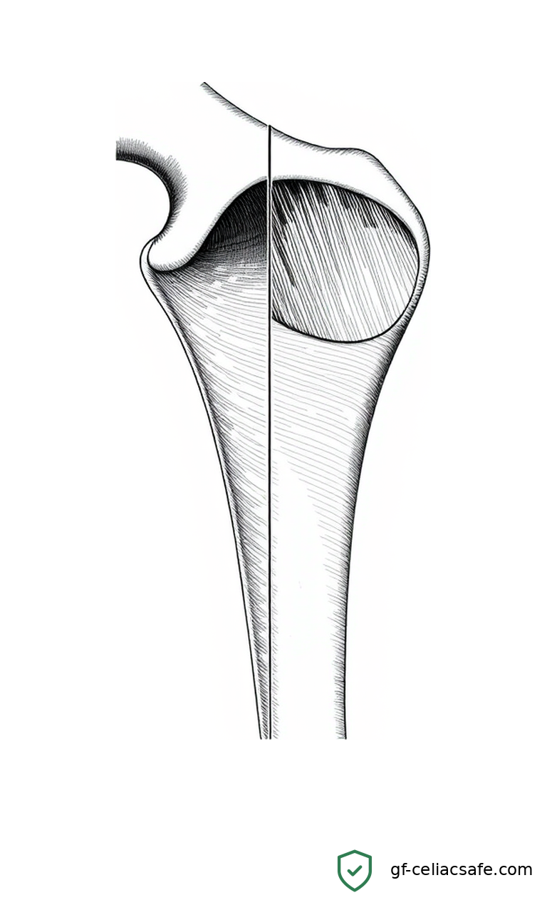
Celiac disease doesn’t just thin bones — it reshapes them. A new multi-center study published in the Journal of Diabetes and Metabolic Disorders found that celiac patients have measurably altered bone geometry at the hip, making their bones structurally weaker in ways a standard bone density scan can’t fully detect.
For celiac families already managing diet, nutrition, and long-term health monitoring, this research adds an important layer: when it comes to bone health, density numbers alone may not tell the complete story.
What This Means for You
The standard test for bone health is a DXA scan — dual-energy X-ray absorptiometry — which measures how much mineral is packed into a given volume of bone. Doctors use this number, called bone mineral density (BMD), to screen for osteoporosis and fracture risk.
This study confirmed what other research has shown: celiac patients have significantly lower BMD at the lumbar spine, total hip, and femoral neck compared to healthy controls. That part isn’t new.
What is worth paying attention to is the finding about bone shape and structure. Celiac patients in the study had thinner cortical bone (the dense outer shell of the bone), less cross-sectional bone area at the hip, and a higher buckling ratio at the femoral shaft and intertrochanteric region (the area where the thigh bone angles into the hip). A higher buckling ratio means the bone is more prone to collapsing under load — like a hollow tube that bends where a solid rod would hold.
In practical terms: the bones of celiac patients were not only less dense, they were also shaped in ways that raise fracture risk beyond what the density number alone would suggest.
For parents raising children with celiac disease, this matters. Bone mass peaks in early adulthood. If chronic inflammation or nutrient malabsorption compromises bone development during childhood and adolescence, that may be difficult to recover. My son has celiac disease, and questions like this — what is celiac doing to his developing skeleton, and are we catching it early enough? — are exactly what I want his care team thinking about proactively, not after a fracture.
The practical takeaway: bone health monitoring for celiac patients should go beyond a single BMD number. Talk to your provider about comprehensive assessment, particularly if nutrient absorption has been poor or diagnosis was delayed.
Key Takeaways
- Celiac patients in this study had significantly lower bone density at the spine, hip, and femoral neck compared to healthy controls.
- Bone geometry was also compromised — thinner outer bone shell, less cross-sectional area, and a higher structural fragility indicator at the hip.
- These geometric changes raise fracture risk beyond what a standard bone density scan predicts.
- One measure — the Trabecular Bone Score — showed no significant difference between celiac patients and controls, suggesting the problem may involve the outer bone shell more than the internal spongy structure.
- Celiac patients should discuss comprehensive bone health monitoring with their care team, especially if diagnosis was delayed or nutrient levels have been low.
The Science
Want to understand how this actually works? We’ll walk you through the technical details below and define every term. No medical degree required.
The Study Design
Researchers at Shiraz University of Medical Sciences enrolled 73 adults with confirmed celiac disease and compared them to 93 healthy controls matched for age, sex, and BMI. All participants underwent DXA scanning — the same imaging used in standard osteoporosis screening — but the team extracted far more information than a typical clinical report includes.
The study examined three distinct measurements:
- Bone Mineral Density (BMD) at the lumbar spine (vertebrae L1–L4), total hip, and femoral neck
- Trabecular Bone Score (TBS) — a software-derived measure of bone microarchitecture, meaning the internal lattice structure of trabecular bone (the spongy tissue inside vertebrae)
- Hip Structural Analysis (HSA) — a geometric analysis of bone shape and cross-sectional properties at three hip regions: the narrow neck, femoral shaft, and intertrochanteric zone
BMD: The Expected Finding
Consistent with prior research, celiac patients showed significantly lower BMD at every measured site. Low BMD for age was more prevalent in the celiac group at the lumbar spine, total hip, and femoral neck.
This connection is well-established. Celiac disease damages the lining of the small intestine, impairing absorption of calcium and vitamin D — the two nutrients most critical to building and maintaining bone mass.
TBS: The Surprising Non-Finding
Here’s where the results get interesting. TBS is designed to measure the microarchitectural quality of trabecular bone — the internal, honeycomb-like structure inside vertebrae. A lower TBS suggests degraded internal bone quality, even when BMD looks acceptable on its own.
In this study, TBS showed no significant difference between celiac patients and controls. On the surface that sounds reassuring, but it also tells researchers something useful: the bone problems in celiac disease appear to affect the cortical (outer shell) bone more than the trabecular (inner spongy) bone. TBS only captures the trabecular lattice; it doesn’t measure what’s happening at the cortex.
Hip Geometry: Where the Findings Get Concerning
The most clinically meaningful results came from Hip Structural Analysis, which extracts geometric properties from DXA scans to characterize bone shape and structural integrity.
Several measures came back significantly worse in celiac patients:
- Buckling Ratio (BR) at the femoral shaft and intertrochanteric region was significantly higher in the celiac group. BR is the ratio of the outer bone radius to cortical thickness — a measure of how susceptible the bone is to buckling under compressive force. A high BR indicates a thinner shell relative to the bone’s width: structurally more fragile.
- Endocortical diameter at the femoral shaft was larger in celiac patients. This sounds like more bone, but it actually means the inner cavity has expanded — so the cortical wall surrounding it is relatively thinner.
Controls outperformed celiac patients on:
- Cortical thickness at the femoral shaft and intertrochanteric region
- Cross-sectional area (CSA) at the femoral shaft — a direct measure of how much bone material is present at that cross-section
Together, these geometric differences describe bones that are not only less dense but structurally compromised at the hip, with thinner walls and a shape more susceptible to fracture under mechanical stress.
Why Geometry Matters Beyond Density
BMD tells you how much mineral is in the bone. Geometry tells you how that bone is shaped and how the mass is distributed. Two people can have identical BMD values but very different fracture risk if their bone geometry differs substantially.
This is why researchers have been pushing to assess bone health in celiac patients using more than density alone. Earlier coverage here explored long-term denosumab treatment for celiac patients with osteoporosis at high fracture risk, which highlighted how complex managing severe bone loss becomes. A study like this one makes the case for earlier, more comprehensive monitoring — before fractures occur.
Related work on body composition changes in girls with celiac disease using bioelectrical impedance analysis showed that the systemic effects of celiac reach beyond the gut in ways that standard clinical metrics often underestimate. Bone geometry is another piece of that picture.
Limitations Worth Noting
This was a cross-sectional study — a snapshot in time — so it can’t establish whether celiac disease caused the geometric changes observed, or track how patients responded to a gluten-free diet over time. The population was based in southern Iran, and disease duration, dietary patterns, and healthcare access may differ from North American or European celiac populations. Longitudinal studies following patients from diagnosis through years of treatment would strengthen these conclusions considerably.
Still, the study adds meaningful evidence that hip structural analysis — not BMD alone — should inform how clinicians assess and monitor fracture risk in celiac disease.
Related Coverage
- Long-term treatment with denosumab in patients with celiac disease and osteoporosis at high risk of fracture
- Bioelectrical Impedance Analysis to Identify the Affected Body Component in Girls With Celiac Disease
References
Naseri A, Keshtkar A, Mofarrah R, et al. “Hip geometry, trabecular bone score, and bone mineral density in patients with celiac disease: a multi-center cross-sectional study in Southern Iran.” Journal of Diabetes and Metabolic Disorders. 2026 Jun 15;25(1):160. https://pubmed.ncbi.nlm.nih.gov/42311855/